Back Of Neck Anatomy ~ Tips For Weekend Warriors Preventing Back And Neck Injuries Baldwin Bone Joint Pc. The superficial lymph nodes of the head and neck receive lymph from the scalp, face and neck. These two ligaments connect and support the spine from the neck to the lower. At the front of the adjacent vertebrae is another connection called the intervertebral disc space. Related posts of muscle anatomy back of neck muscle anatomy labeling. It runs down the back part of the neck, and opens into the external jugular vein just below the middle of its.
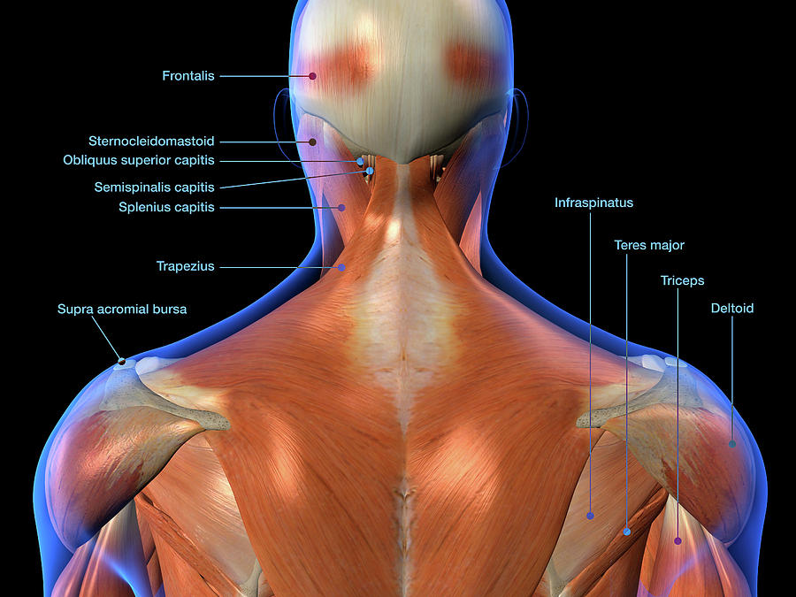
The sternocleidomastoid divides the neck into anterior and posterior triangles. The neurocranium (cranial vault) and the viscerocranium (facial skeleton). The neck is essentially a passageway for air, food, liquids, blood, and more to travel between the head and the rest of the body, through structures such as blood vessels, nerves, and lymph nodes, as well as the larynx, trachea, and esophagus. In the cervical spine, the spinal cord connects to the brain at the base of the skull. The anterior triangle of the neck is made by the anterior border of the sternocleidomastoid muscle, the inferior border of the mandible and the midline of the neck.
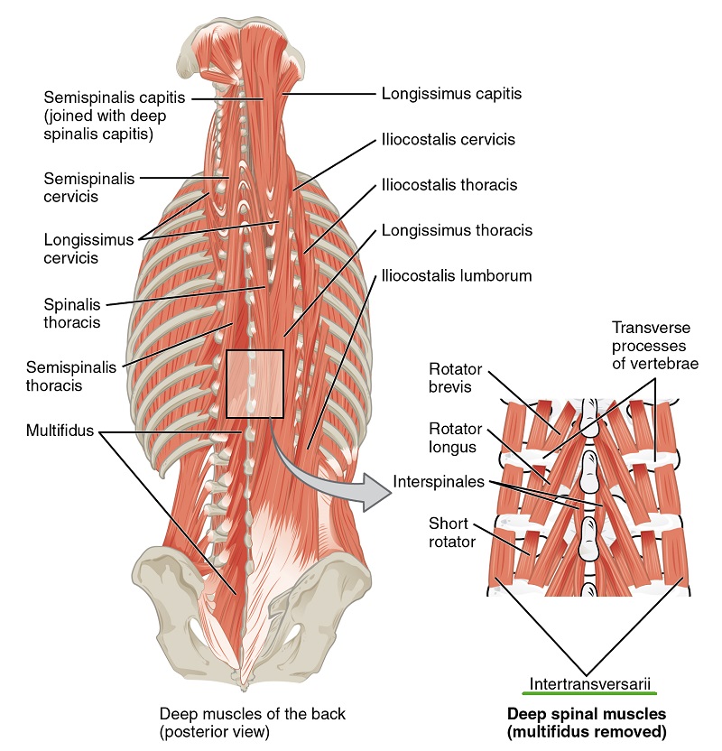
Topographic Anatomy Of The Back The Lecturio Medical Online Library from d3uigcfkiiww0g.cloudfront.net The nerves of the head and neck include the most vital and important organs of the nervous system — the brain and spinal cord — as well as the organs of the special senses. The posterior external jugular vein (v. The neurocranium (cranial vault) and the viscerocranium (facial skeleton). In addition, in this region we also find the major cranial and spinal nerves that connect the central nervous system to the organs, skin, and muscles of the head and neck. The internal jugular vein commences at the jugular foramen, and is the direct continuation of the sigmoid sinus, which is a large vein draining blood from the vein. The neck is a vital part of the human body. The occipital bone surrounds a large opening known as the foramen magnum. The crossword solver found 20 answers to the back of the neck crossword clue.
The vertebral bodies are round shapes.
They empty into the right and left subclavian veins in the base of the neck. The majority of these nerves control the functions of the upper extremities and allow you to feel your arms, shoulder, and back of your head. Enter the answer length or the answer pattern to get better results. It runs down the back part of the neck, and opens into the external jugular vein just below the middle of its. The largest vein in the neck is usually the internal jugular vein, which drains blood from the brain, neck muscles, face and organs of the neck. Extending from underneath the chin, to the posterior aspect of the head. This triangle can be further divided into the submandibular triangle, submental triangle, muscular triangle and carotid triangle. The muscles of the back and neck that move the vertebral column are complex, overlapping, and can be divided into five groups. 12 photos of the anatomy of the back of the neck anatomy of back of human neck, anatomy of the back and neck, anatomy of the back of the neck, anatomy of the back of the neck muscles, anatomy of the back of your neck, human anatomy, anatomy of back of human neck, anatomy of the back and neck, anatomy of the back of the neck, anatomy of the back of the neck muscles, anatomy of the back of your neck These muscles give the sides of the neck their. In the neck are the thyroid and parathyroid glands, that secrete hormones that control metabolism and blood calcium levels. The posterior external jugular vein (v. The top of the cervical spine connects to the skull, and the bottom connects to the upper back at about shoulder level.
They empty into the right and left subclavian veins in the base of the neck. The occipital bone surrounds a large opening known as the foramen magnum. In the neck are the thyroid and parathyroid glands, that secrete hormones that control metabolism and blood calcium levels. Inside the facet joint is synovial fluid, which lubricates the joints. The external jugular veins descend on either side of the neck, passing over the sternomastoid muscles and beneath the platysma.
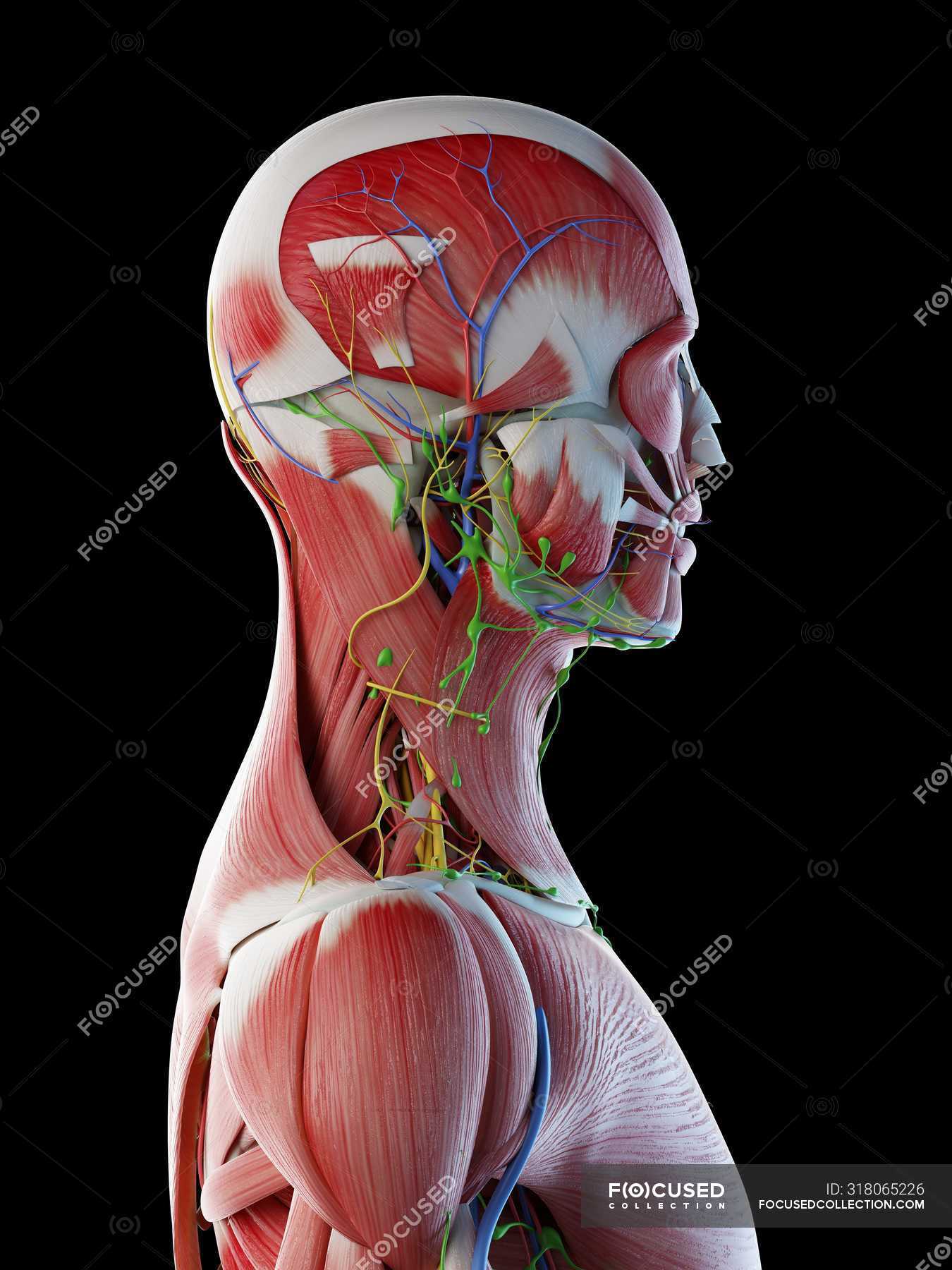
Labeled Anatomy Chart Of Neck And Back Photograph By Hank Grebe from images.fineartamerica.com In addition, in this region we also find the major cranial and spinal nerves that connect the central nervous system to the organs, skin, and muscles of the head and neck. It consists of two major parts: The four parathyroid glands are situated upon the back surface of the thyroid gland. The occipital bone surrounds a large opening known as the foramen magnum. The occipital bone is a bone that covers the back of your head; This is a more stylized study and not meant to be entirely cor. The cervical spine supports the weight and movement of your head and protects the nerves exiting your brain. 12 photos of the anatomy of the back of the neck anatomy of back of human neck, anatomy of the back and neck, anatomy of the back of the neck, anatomy of the back of the neck muscles, anatomy of the back of your neck, human anatomy, anatomy of back of human neck, anatomy of the back and neck, anatomy of the back of the neck, anatomy of the back of the neck muscles, anatomy of the back of your neck
The cervical spine supports the weight and movement of your head and protects the nerves exiting your brain.
Two of the main ligaments in the back are the anterior longitudinal ligament and the posterior longitudinal ligament. The muscles of the neck stabilize and move the head. Remember, while c1 and c2 allow tremendous ranges of neck movement, they are supporting your head too. It consists of two major parts: It contains the cervical spinal cord, muscles, and major blood vessels that supply the entire body. In addition, in this region we also find the major cranial and spinal nerves that connect the central nervous system to the organs, skin, and muscles of the head and neck. Enter the answer length or the answer pattern to get better results. Nerve roots exit the spinal cord in the neck and provide control and sensation to different parts of the body. The posterior external jugular vein (v. They are arranged in a ring shape; In the neck are the thyroid and parathyroid glands, that secrete hormones that control metabolism and blood calcium levels. The sternocleidomastoid divides the neck into anterior and posterior triangles. The neurocranium (cranial vault) and the viscerocranium (facial skeleton).
They empty into the right and left subclavian veins in the base of the neck. The internal jugular veins form the major venous drainage of the head and neck and are deep veins that parallel the common carotid artery. The occipital bone surrounds a large opening known as the foramen magnum. These muscles give the sides of the neck their. Each nerve provides sensation to a specific area of the body called a dermatome.
Male Anatomy Of Head Neck And Back With Musculature Computer Illustration Normal Transparent Stock Photo 318065226 from st.focusedcollection.com The cervical spine supports the weight and movement of your head and protects the nerves exiting your brain. Remember, while c1 and c2 allow tremendous ranges of neck movement, they are supporting your head too. Nerve roots exit the spinal cord in the neck and provide control and sensation to different parts of the body. They are arranged in a ring shape; It runs down the back part of the neck, and opens into the external jugular vein just below the middle of its. They ultimately drain into the deep lymph nodes. The back of the neck is mostly comprised of muscles, as well as the spine. It provides support and mobility for the head.
They ultimately drain into the deep lymph nodes.
An area called the occiput. The facet joints in the neck are where the back of adjacent vertebrae join together. They ultimately drain into the deep lymph nodes. The top of the cervical spine connects to the skull, and the bottom connects to the upper back at about shoulder level. In the neck are the thyroid and parathyroid glands, that secrete hormones that control metabolism and blood calcium levels. Extending from underneath the chin, to the posterior aspect of the head. The sternocleidomastoid divides the neck into anterior and posterior triangles. The neck is a vital part of the human body. The posterior external jugular vein (v. It contains the cervical spinal cord, muscles, and major blood vessels that supply the entire body. The neck is essentially a passageway for air, food, liquids, blood, and more to travel between the head and the rest of the body, through structures such as blood vessels, nerves, and lymph nodes, as well as the larynx, trachea, and esophagus. This is a more stylized study and not meant to be entirely cor. Two of the main ligaments in the back are the anterior longitudinal ligament and the posterior longitudinal ligament.
Share :
Post a Comment
for "Back Of Neck Anatomy ~ Tips For Weekend Warriors Preventing Back And Neck Injuries Baldwin Bone Joint Pc"

Post a Comment for "Back Of Neck Anatomy ~ Tips For Weekend Warriors Preventing Back And Neck Injuries Baldwin Bone Joint Pc"